Conduction System of the Heart
One of the more unique abilities of the cells within the heart is automaticity, the ability to create its own electrical activity, and inherent rhytmical electical activity is what allows a heart to contract with some level of rhythm. This conduction process is developed during early embryonic stages of development, in which as little as 1% of the cardiac cells devellop autorhymicity. This 1 % goes on to develop the primary pacemaker sites that form to make the conduction system of the heart.
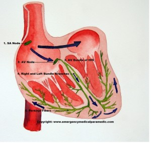
The following is an image of the normal conduction system of the heart:
(1.) The SA node is the first to fire off the intrinsic, rhythmic electrical current (that later causes the atrium and ventricles to contract). 2. This current then travels through the right atrium and triggers the AV Node (2.) to continue the conduction down the AV Bundle of HIS (3.), further down into the Left and Right Bundle Branches (4). and finally into the large branches of the Purkinje Fibers(5.).
Here is a video outlining the conduction of system of the heart:
httpv://www.youtube.com/watch?v=te_SY3MeWys
Conduction Pathway of the Heart
1. The SA Node has autorhymic fibers that are capable of initiating cardiac action potentials and setting the basic pace for the heart.
2. The Atrioventricular Node receives the action potentials from the SA Node and triggers the conduction of the Atrioventricular Node Bundle of HIS.
3. The Atrioventricular Node Bundle of HIS passes the action potentials (conduction of eletricity) through the left and right Bundle Branches.
4. The Left and Right Bundle Branches recieves action potentials from the AV Bundle of HIS and passes them onto the Purkinje Fibers.
5. The Purkinje Fibers receives the action potentials and passess them to the ventricular myocardium, which is the muscle responsible for the ventricle contraction and the mechanical pumping of the heart.
Return to: ECG Interpretation Tutorial.
Next page in the ECG Interpretation Tutorial:

