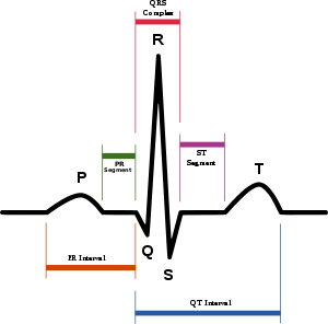
Normal ECG
Normal ECG Components. As a paramedic, given that we generally treat the elderly and persons with known cardiac disease or a multitude of medical diseases and disorders, it is very unlikely to see a perfectly normal ECG. In reality, even with younger people and genuinely healthy people, it is uncommon to see a perfect, normal ECG. But, you need a baseline in which to work with, so this page provides information about the mythical “perfectly normal ECG.”
Normal Components of an ECG
(Image Source: Wikipedia 2011)
P Wave
Normally precedes the QRS complex and is upright in leads: I, II, aVF, and V4 and V5. Inverted (negative) in leads II, aVL and V1 and V2. Duration of P wave is usually under 0.1 Second. Amplitude should be 0.5-2.5 mm when viewed in lead II. The normal shape of the P wave is smooth and rounded.
QRS Complexes
Regularly follows a P wave at regular intervals. 0.10 seconds or less in duration. Shape generally narrow with sharply pointed waves.
T Waves
Amplitude less than 5mm in the standard limb leads and less than 10 mm in the precordial leads.
PR Interval
0.12 seconds -0.20 seconds.
QT Intervals
Less than half the preceding R-R intervals
ST Segments
Normally flat, but may commonly be depressed or elevated but no more than 1.0 mm.
Normal ECG Patient Presentation
A patient with a normal ECG, given no other health concerns, should present well perfused with skin that is warm, pink, dry, their level of consciousness should be normal, their blood pressure may or may not be normal, and their pulse rate should be between 60 and a 100 per minute in a fundamentally regular and strong rythym.
Return to: ECG Interpretation Tutorial.
Next page in the ECG Interpretation Tutorial:


